Neurology - CVAs
*Types of Stroke
- 80% are Ischemic infarct stroke
- ONLY 20% are Hemorrhagic stroke
*Ischemic Stoke
*Hemorrhagic Stroke
- 2 basic AREAS with their own subtypes of ruptures
--> SAH = congenital Berry aneurism ruptures
--> intraparenchymal DEEP ruptures of C-B aneurisms of LSA and CAA angiopathy
*CVA MAP
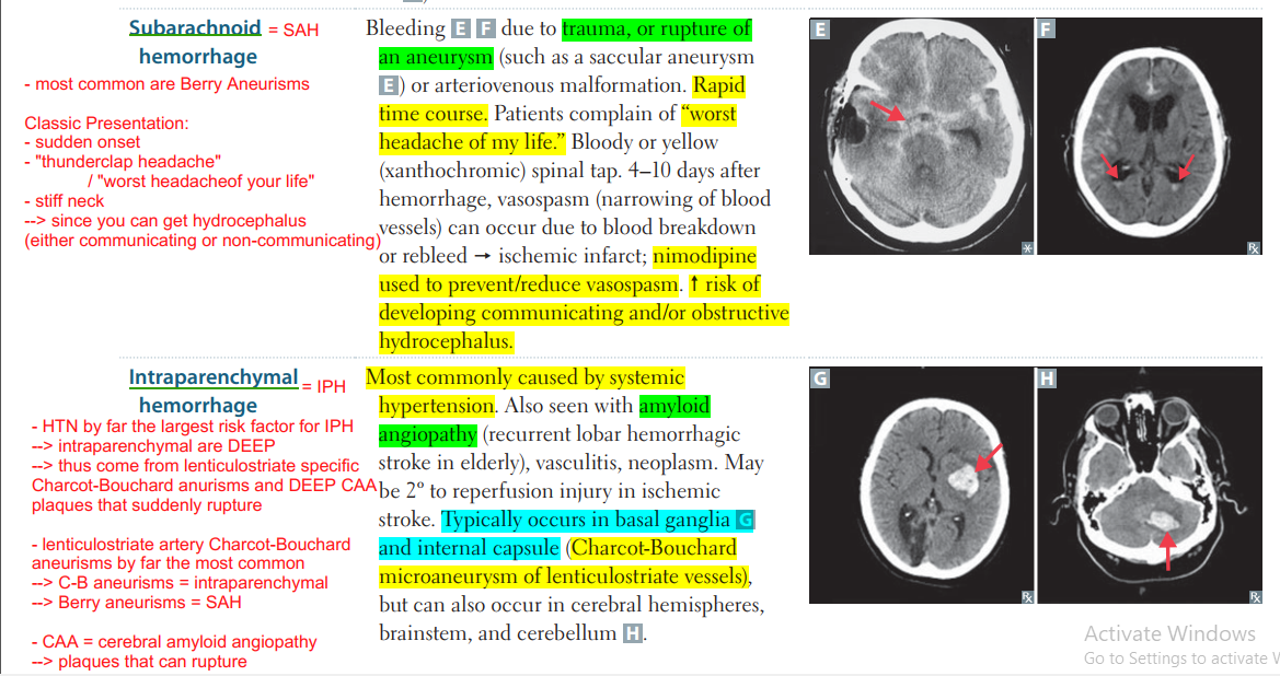
*SAH - Hemorrhagic Stroke
- SAH = congenital Berry aneurism ruptures
-->
*Intraparenchymal DEEP - Hemorrhagic Stroke
- 2 basic ruptures in IPH
- ruptures of Charcot-Bouchard anurisms of lenticulostriate artery
- DEEP CAA = cerebral amyloid angiopathy
plaques that suddenly rupture
*CAA = Cerebral Amyloid Angiopathy - IPH Strokes
- CAA are most commonly found in the frontal and parietal lobes
--> but they most commonly cause strokes in the parietal and occipital lobes - parietal (34.1%) and occipital (29.3%) regions
--> they NEVER occur in the white matter, BG, or brainstem
*Brain Aneurisms
- rupture causes 2 types of hemorrhage depending on location of the aneurism
--> SAH = congenital Berry aneurism ruptures
--> intraparenchymal DEEP ruptures = C-B aneurisms
*Berry Aneurisms
- rupture causes SAH - Hemorrhagic Stroke
*Charcot-Bouchard Aneurisms
- rupture causes IPH - Hemorrhagic Stroke
*Recovery of Stroke timeline
- different stages of inflammatory cells in the brain repairing damage from stroke
--> days <1, 1-3, 3-5, 1 week, 2 weeks - eosinophils, then neutraphils, then macrophages
--> week 1 = gliosis and vascular proliferation
--> week 2 = glial scars fully formed
*Lacunar Stroke
- biggest risk factors = uncontrolled HTN and diabetes
--> smoking also - most common type of ISCHEMIC stroke
- "lacunar" refers to LAKES of fluid that form after cells have been damaged
--> these lakes are from ischemia led necrosis and edema - most are in the second segment of the middle cerebral artery
--> deep in the brain at the basal ganglia
--> most common = lenticulostriate artery to BG
*Global Ischemia
- Global Ischemia affects illegally parked cars FIRST
- "CAN'T PARK NEAR here"
--> Caudate
--> Perkinje of Cerebellum
--> neocortex
3 causes of Lacunar Stroke
- all are due to chronic HTN --> by far most important
--> secondary risk factors are diabetes and smoking - lipohyalinosis (thickenning)
- microatheroma formation,
- hardening/thickening of the vessel wall
--> = (hypertensive arteriolar sclerosis)
*Home
*Home
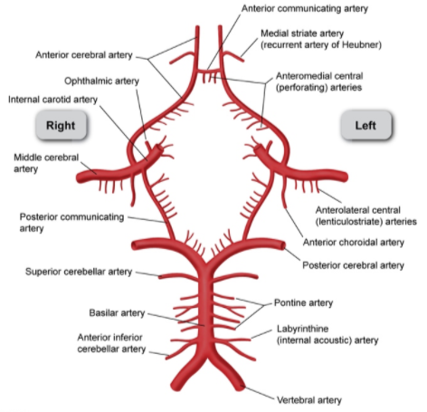
*Internal Carotid Sinus Emboli
- go up to the MCA and ACA of Circle of WIllis
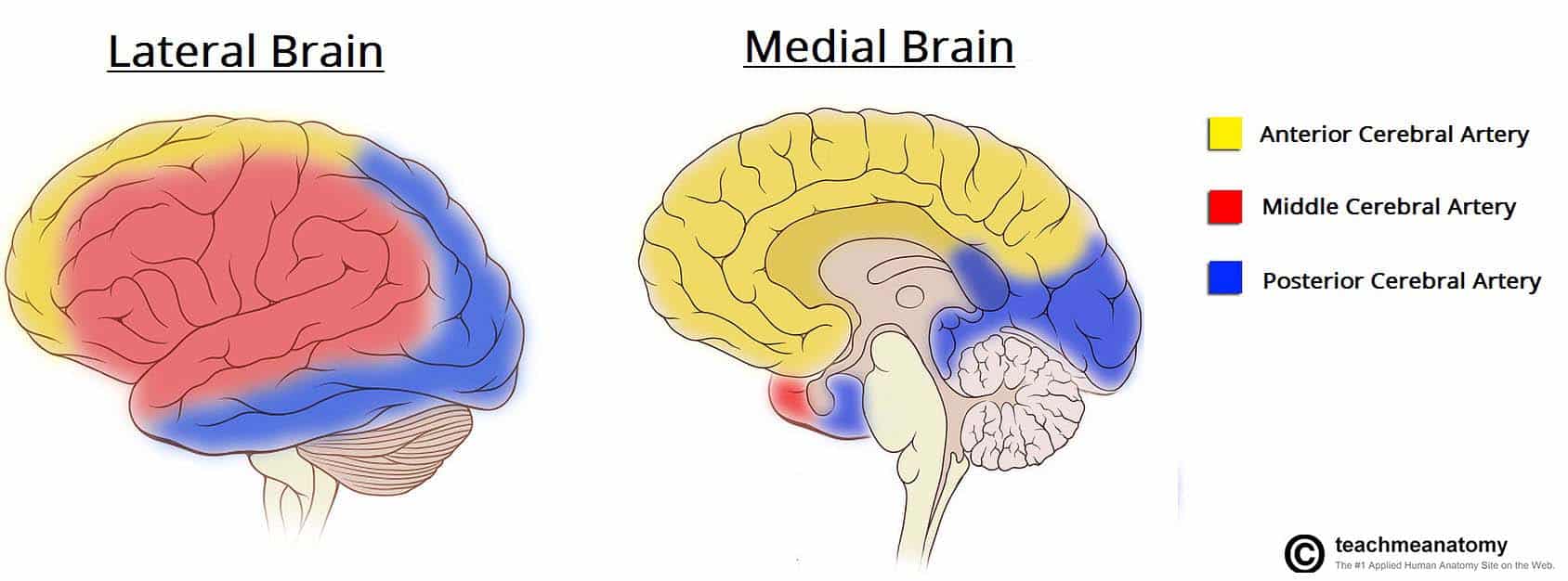
*MCA
- caontains the lenticulostriate artery
- Brain Blood supply: supplies upperlimbs and face of the HUMNCULUS
--> upper limbs and face
* lenticulostriate artery
- most common artery affected by HTN and diabetes infarct CVAs
- hit the basal ganglia, usually putamen
*Putamen lenticulostriate CVA
- usually from Charcot-Bouchard aneurisms from chronic HTN, diabetes and smoking
Putamen hemorrhage CT
Left Atrial Appendage Emboli
- A Fib
*ACA
- caontains the Anterior communicating artery
- Brain Blood supply: supplies the middle sulcus of the HUMNCULUS
--> lower limbs up to pelvis
Basilar Artery
- runs in front and under the PONS
- gives off the Pontine artery branches
*Pontine artery branches
- supply the PONS
.
.
.
.
.
.
.
Vertbral Artery Branches
- IACA and anterior spinal arteries
Vertbral Artery Branches
- IACA and anterior spinal arteries
*Anterior Spinal Artery Lesion
- ALC = descending corticospinal tracts
--> voluntary motor control - ALS spinothal
--> A = sensation for pain and temp
--> L = sensation for pain and temp
Vertbral Artery Branches
- IACA and anterior spinal arteries
*Anterior Spinal Artery Lesion
- ALC = descending corticospinal tracts
--> voluntary motor control - ALS spinothal
--> A = sensation for pain and temp
--> L = sensation for pain and temp
click to edit
*PONS
- lesions and bleeds present with PONS = PINPOINT PUPILS
- rapidly evolving coma
- loss of horizontal gaze
- quadraparesis
*DIRTY USMLE - CVAs
DIFFUSE on ONE SIDE = Brainstem Stroke
Midbrain
PONS
Medulla
*Brainstem CVAs
Rule of 4s
- 4 CN rule = MIDNIGHT 12 MIDLINE CN
- 4 MEDIALs
--> 2 MEDIAL GENERAL MOTOR (CorticoSPINE + corticoBULBAR CN)
--> 2 MEDIAL specific (MEDIAL LF + MEDIAL Lemniscus)
*Rule of 4s
-
4 MAINS = MEDIAL
Rule of 4
2 MEDIAL GENERAL MOTOR (CorticoSPINE + corticoBULBAR CN)
- MOTOR nuclei of CN cranial nerves
--> all the groups from CN MIDNIGHT 12 = MIDLINE
--> (above RULE of $ for CNs) - MEDIAL /midline CORTICOspinal tract
--> main muscle tracts
--> everything below it will be affected
2 MEDIAL specific (MEDIAL LF + MEDIAL Lemniscus)
- MEDIAL lemniscus
--> vibration and proprioception - MLF = medial Longitudinal Fasciculus
--> seen in MLF INO with MS SIN
4 SENSORY = SIDE
Rule of 4
SENSORY Syndromes are to the SIDE = lateral
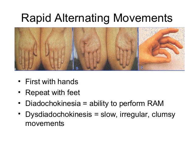
RAM = rapid alternating Movement
- dysdiadocokinesia
Symathetic Chain = Horners syndrome
- Horners triad
--> miosis
--> anhydrosis
--> ptosis
4 CN SECTIONS
*MIDNIGHT CN 12 = MIDLINE
Rule of 4
Numbers that DIVIDE 12 = MIDLINE
Clinical Cases
Clinical Case
Clinical Case
Notes:
- note that
Clinical Case
Notes:
- note that
Clinical Case
DIFFUSE on ONE SIDE = Brainstem Stroke
Midbrain
PONS
Medulla
*Brainstem CVAs
Rule of 4s
- 4 CN rule = MIDNIGHT 12 MIDLINE CN
- 4 MEDIALs
--> 2 MEDIAL GENERAL MOTOR (CorticoSPINE + corticoBULBAR CN)
--> 2 MEDIAL specific (MEDIAL LF + MEDIAL Lemniscus)
Clinical Cases
Clinical Case
Clinical Case
Notes:
- note that


