DNA and Cell Structures
Junctions between Cells
Cadherins
Notes:
- cadherins are the proteins and adhesion molecules that connect adjacent epithelial cells at the points of desmosomes
--> form the anchor between adjacent epithelial cells - note that cadherin proteins need Ca++ in order for them to bind each other
--> think cadherin = Calcium - intracellularly cadherins attach to desmosomes of cells
- extracellularly cadherins bind to other cadherins of other epithelial cells with the help of calcium
DNA replication and
Protein Synthesis
Transcription
- transcribe DNA --> mRNA
Post-transcriptional modifications of RNA
--> hnRNA --> pre-mRNA --> mature mRNA
- 3 major steps of postranscription modification
- 5' Cap
- 3' Poly A Tail
- Splicing out of introns
--> splicosome = collection of snRPs and proteins
post-transcriptional modification
Genetic *Mutations
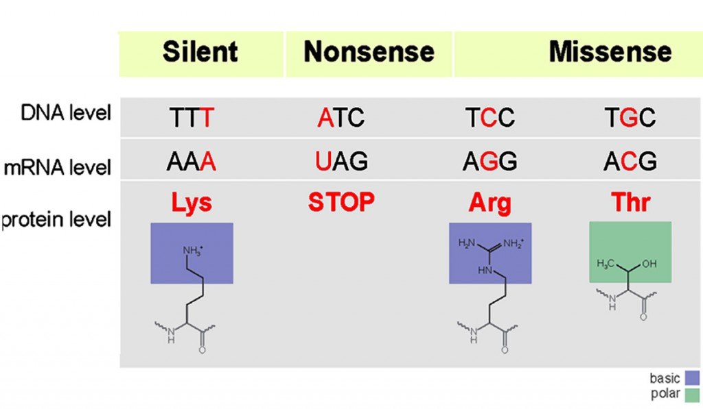
Genetic Mutation Types
*Missense mutations
-
*Nonsense mutations
- missense mutation that actually inserts a STOP codon
--> early breakage of the protein
*Frameshift mutations
- note always check for not having MULTIPLES of 3
--> if there is a multiple of 3 added, then you CANNOT have a frameshift mutation
*Silent mutations
- mutations that only have a point mutation in an AA
- they lead to NO change in the AA inserted
Allelic Heterogeneity
Notes:
- note that whenever talking about ___ heterogeneity, this means there is difference = hetero- within that object that results in the same phenotype
--> the exception is phenotypic heterogeneity ofcourse where it means different phenotypes from the different mutations on the same gene - in allelic heterogeneity we are talking about possibly different mutations within the same allele, but they all manifest as the same phenotype
- in genetic heterogeneity we are talking about possibly different gene mutations within different genes, but they all manifest as the same phenotype
- in phenotypic heterogeneity we are talking about different phenotypes arising from the same gene undergoing different mutations
- note in the above that polygenic disease means that a disease is cause by defects in multiple other genes
--> best example is T2 DM which is clearly hereditary, but so many different genes are involved that we don't know of
Case example:
Insulin
Preproinsulin and
Insulin Generation
Notes:
- note that preproinsulin has an N-terminal signaling sequence that is read to direct it to the rough ER for final translation
- translation of the preproinsulin mRNA actually starts in the cytosol as the mRNA is moving out from the nucleus
- the preproinsulin mRNA is being translated in the cytosol when the DNA Pol A reaches the signal sequence at the N-terminus
--> this immediately stops the translation and signals for SRPs = signal recognition particles to transport it to the rough ER for further translation - preproinsulin then cleaved into proinsulin, which forms loop structure inside the rough ER
--> made of A and B chain polypeptides and with a C-peptide loop at the end - sent to golgi in transport vessicle, further packaging in the Golgi and again buds off into a secretory vesicle
- once in a secretory vessicle and heading towards the plasma membrane for transport outside the cell, the C-peptide is cleaved from the proinsulin molecule
--> in the secretory vessicle you now have C-peptide and insulin that are released from the cell and into circulation
Example:
*Lab Techniques
Radiolabelled Assays
- fix antibodies to an assay
- put antigens in that are radiolabelled
- meaasure the radioactivity to see how much antigen is bound to the antibodies
- can be used to compare whether proteins/antigens have common epitopes
Example:
Notes:
- in this case the antigens X and Y are put into the same assay
- Antigen X is added and creates radioactivity
- since the radioactivity stays the same as Antigen Y is added, this means that it does NOT bind to the assay and hence has no common epitopes as Antigen X does
Notes:
- 3 main post-transcriptional modifications
- 5' methyl-guanosine capping
--> helps stabilize the 5' end of mRNA from being degraded in the cytosol - 3' Poly-A tail capping
--> helps stabilize the 3' end of mRNA from being degraded in the cytosol - removal of introns from the mRNA by snRNPs = small nuclear ribonucleotide proteins
--> exons are "actual DNA", "introns are out"
Poly A tail and complement
Notes:
- note always think of the 3 major post transcriptional changes to mRNA and their complements
Tight Junctions
Blood Brain Barrier
- Blood BB is possible through Tight Junctions
Blood BB and Tight Junctions
Clinical Case
Notes:
- note the main reason why the Blood BB is able to keep molecules out is through tight junctions
- these tight junctions are also supported by astrocytes cells that surround the cappilaries
- there are no fenestrations = opennings in the Blood BB capillaries either
Gap Junctions
Notes:
- note that in preparation for delivery nearing the end of a pregnancy, myometrial cells start to express connexin proteins and oxytocin
- connexin proteins are needed in the myometrium for gap junctions betweent he muscle cells so they can forcefully contract
- oxytocin receptors are needed since oxytocin from the posterior pituitary gland initiates contractions
Case example:
Hemi's AdDES integrin to ANCHORS
- anchor junctions are made by:
Adherin junctions --> cadherin
desmosomes --> cadherins + others
hemidesmosomes --> integrin
connexin CONNEX the GAPS
gap junction --> connexin
Tight junctions OCCLUDE water soluble drugs from the Blood BB
tight junctions --> occludin and claudins
Cellular Proteins / Structures
Cell transport
- vessicles
- microtubules
- kinesein and dyneins
*Organelles
Microtubules
Kinesins and Dyneins
Kinesin Transport Example
Clinical Case
Notes:
- note that kinesins transport secretory vessicles towards the growing end = positive end of microtubules = AWAY from the centre of the cell and towards the periphery
--> classic example would be ACh NTs that are brought to loading zones at the cell membrane near Ca++ channels to be ready for release
"DYNesins DINE with MINE at HOME,
Kinesins are KIND and POSITIVE and dine away"
- Dynesins are proteins that transport vessicles along microtubules
--> towards negative (-) end
--> towards the cell centre - Kinesins are proteins that transport vessicles along microtubules
--> towards positive + end
--> towards the periphery
Protein Synthesis Codons Overview
- reading DNA
- reading mRNA
- building AA with tRNA
Start and Stop Codons
START Codons: Are U Going? (to start?)
- AUG
STOP Codons: U Are Annoying!! ... U Go Away!! ... U Are Gone!!
- UAA
- UGA
- UAG
DNA Structure
*Telomeres
- telomerase adds TTAGGG repeats at the 3' ends of DNA
--> these end pieces = TAGs are telomeres - prevent them from being degraded
- this is mostly done in the stem cells of the body since they need much longer telomeres
- other mature cells of the body have shorter telomeres
--> when a telomere reaches a certain short length it signals for apoptosis of that cell
*Collagen Protein
- most abundant protein in the human body
- major component of connective tissue + extracellular matrix
- 4 subtypes of collagen
4 Types of Collagen
- to remember 4 types of collagen --> be COLLEGIATE and spell them out
type 1 --> ONE --> bONE
--> bone, tendons, skin
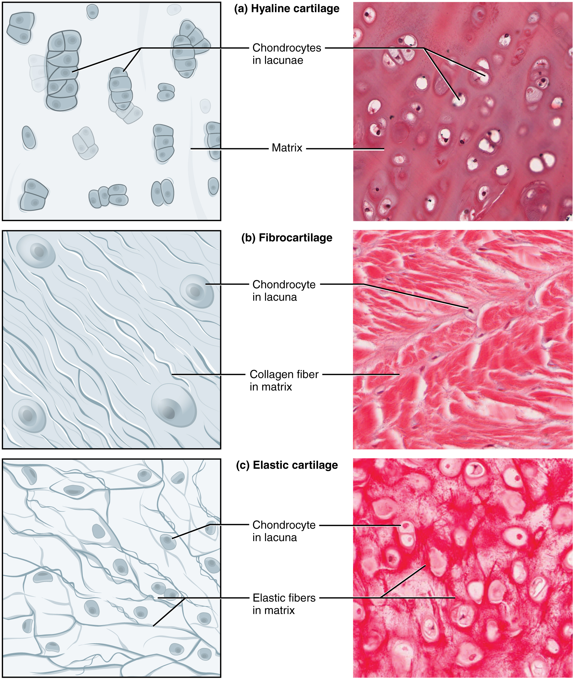
type 2 --> TWO --> carTWOlage
--> type 2 collagen makes up cartilage
--> especially hyaline cartilage
type 3 --> 3 D --> ThreE D --> Ehlor Danslos and others
--> Ehlor Danslos affect synthesis of type 3 collagen
--> blood vessels, also uterus and fetal tissue
--> NOTE that MARFANS is NOT COLLAGEN like Ehlors even though they have similar presentations
--> MARFANS is extracellular fibrillin scaffolding for elastin
type 4 --> Four --> FLOOR = basement membrane
--> any disease with basement membrane
--> GBM = Goodpasture Syndrome with antibodies to the renal GBM and pulmonary BM
MI and collagen 1 deposition scarring post MI
Notes:
- note the period 2 wks --> 2 months post MI your heart starts laying down fibrotic scar tissue on the damage myocytes of the heart
- recall that myocytes can't regenerate so your body has to replace them with collagen type 1 scarring
- type 1 collagen is the same that is in bones, tendons, think the starting materials for tissue
Example:
Cartilage
Skeletal Muscle
*Sarcomere Structures
- see other note under musculoskeletal
SNoW DRoP BLOT tests
- DNA
- RNA
- Proteins
Western Blot
- protein identification
Western Blot example 1
Notes:
Example:
Amino Acids and their Derivatives
Arginine
- Arginine makes NO through eNOS
- eNOS = endothelial NO synthase
- responsible for vasodilation of blood vessels in response to
--> Bradykinin, sub p, etc.
Arginine --> NO synthesis --> vasodilation
Notes:
- note that
Clinical Case
Tyrosine
- used to make Monoamines
--> Tyrant wants MONO control
*Types of Receptors
- receptors are broadly either cell surface receptors or intracellular receptors
- note MOST horomones are lipophillic and bind inside the cell
--> exceptions
Intra vs Outer Cell receptors
Intra vs Outer Cell receptors - Hormones example
Case presentation:
Notes:
- note that most hormones are lipophilic and bind inside the cell
- think of hormones as being POWERFUL, but they take time for them to work and LASTING
--> hence bind INSIDE the cell
--> transcription changes genes - things like insulin and glycogen bind outside the cell
Notes:
- most horomes bind intracellular
- Exceptions of Hormones that bind outside the cell:
- GH = Growth Hormone
--> JAK-STAT pathway
--> think of Silver fox teacher - parathyroid hormone = PTH
--> think PTH has tight control on calcium so has to be quick and bind outside
Histone Proteins
- recall nucleosome cores have 8 proteins
- H2a, H2b, H3, H4 x 2
- H1s are are on their own = 1
--> outside the core, H1 holds together linker DNA
Telomeres and telomerase in Malignancy
Case example:
Notes:
- note
*RNA
- subtypes
- location made, etc.
Synthesis and Function of 3 main RNA subtypes
- remember RNA 123
- RNA subbtypes RNA 123 makes RMA
R / 1 = RNA 1 make ribosomes
M / 2 = RNA 2 makes mRNA
A / 3 = RNA 3 make the AA builder = tRNA
RNA case
- note that ribosomal rRNA is made and assembled in the nucleolus
- note that basophilic means "stains dark"
--> how they described nucleolus
Notes:
- note that
Clinical Case
Western Blot example 1
Example:
Notes:
*Translation by rRNA and tRNA
- mRNA to polypeptides (amino acid sequences --> folded into proteins)
transfer RNA = *tRNA
- carries AAs tot he ribosome to add to the polypeptide chain
ribosomal RNA = *rRNA
- 60S and 40S
- releasing factors bind to the ribosome and casue the release of the polypeptide when the mRNA reaches one of the 3 STOP codons
--> UAA
--> UGA
--> UAG
*Ribosomes - Cytosol Ribosomes vs. RER Ribosomes
- RER ribosomes
--> send proteins for cell membrane, secretory, nucleus (NOT the nucloelus), Golgi and lysosomes
Modified Translation during Apoptosis
- use of the IRES = internal ribosomal entry site
- this starts translation
- instead of the normal recognition of the 5'prime cap
Notes:
- note that in apoptosis, can't use the normal recognition of the 5'prime cap
- in normal translation, the small unit of the ribosome binds to the 5' cap
- the 5' cap is found by eIFs = eukaryotic initiation factors
- instead during apoptosis, these eIFs are degraded and need to use the IRES = internal ribosomal entry site to start translation
- note also here that normally during eukaryotic translation, there are not multiple open reading frames
--> there are multiple reading frames in prokaryotes, but NOT eukaryotes
--> not sure why I thought this was true for eukaryotes?
Clinical Case
Reading mRNA: Translation
- mRNA read in the 5' to 3' direction
- opposite to DNA reading
Reading mRNA Practical Example
- mRNA is read in the 5' --> 3' direction
- opposite to DNA reading
- think the AAs need to match the original DNA so the mRNA is read in the opposite direction to the DNA
Notes:
- here first look for the stop codons
- remember to read mRNA in the 5' to 3' direction
- find UAA UGA UAG
- then it is asking for the codon before this
- make sure to put the 3' and 5' in the right place, they switch them sometimes
Clinical Case
Enzymes
- Helicase = stairCASE unwinder
- ssBPs = single strand binding proteins
--> holds the unwound DNA open - gyrase = topoisomerase II = stops supercoiling
- RNA Primase = lays the initial primer for the DNA Pol 3 on both leading and lagging strands
--> note that RNA primase is the ONLY enzyme in DNA replication that makes RNA and not DNA
--> doesn't need to be DNA as it is removed by DNA Pol 1 anyways - DNA Pol III = think DNA read in 3' --> 5' direction = DNA POL 3 at
--> starts after primase lays first
--> reads DNA in 3' --> 5'
--> makes DNA in 5' --> 3' and proof reading 3' --> 5'
--> proof reads in BACKWARDS direction ONLY
--> can't take out bits and add at the same time like DNA POL 1 - DNA Pol 1 = the ONE DNA Pol that has both 5' --> 3' synthesis and exonuclease in the same direction
(DNA pol 3 ONLY has backwards = 3'-->5' exonuclease backwards proof reading)
Pre Replication Fork Factors for Transcription
- Transcription Enhancers = several base pair upstream ? how many
- CAT TATA TAT = initiating and TATA box promotor base pair upstream ? how many
--> right before the genes to be encoded
Transcription Enhancer
- located even more upstream from the CAAT initiater sequence
- creates a loop in the DNA after DNA POL has started and makes it go faster
CAAT and TATA box
- Song = "CATS comin down the track... "
.- "TATA TAT" - CAAT = initiating sequence for DNA transcription
- TATA box = promoter region after CAAT
CAAT and TATA box example
Notes:
- note that
Clinical Case
DNA Coding Strand = Sense strand
vs Template Strand
Notes:
- coding strand = sense strand
--> identical to mRNA - template strand
--> is the template for RNA to copy as a template
Practical Transcription - Practise Qs
- coding strand / sense strand = actual DNA that is the same as the mRNA made
--> remember the protein is the important thing and ultimate goal
unwinder - template strand / nonsense strand = template DNA for the mRNA to be made from
--> think it would not make the protein
--> it is nonsense
Notes:
- note that the exonuclease and DNA/mRNA synthesis 5' --> 3' always refers to the strand being made
- ex - DNA is read in the 3' --> 5' direction, so DNA Pol 3 has 5' --> 3' direction synthesis
COLL A GEN synthesis
- "COLL A GEN" = CAG rule of 3s
- C = Vitamin C / Scurvy needed in collagen synthesis
- A = alpha chains = 3 of them
- G = GLYCENE rule of 3 in collagen
- Collagen Rule of 3s = 1/3 AAs in collagen are GLYCINE
*Home
DNA Damage and *Repair
Base Excision Repair
- GEL PLease
- glycosylase , endonuclease, lyase, polymerase, lygase
- this is usually for switching out wrong individual bases, etc
--> not quite the same as dimers in UV damage
*Modifications of DNA
- methylation
--> CpG = MUTED
--> Histones = MAY be MUTED - acetylation = ACTIVE
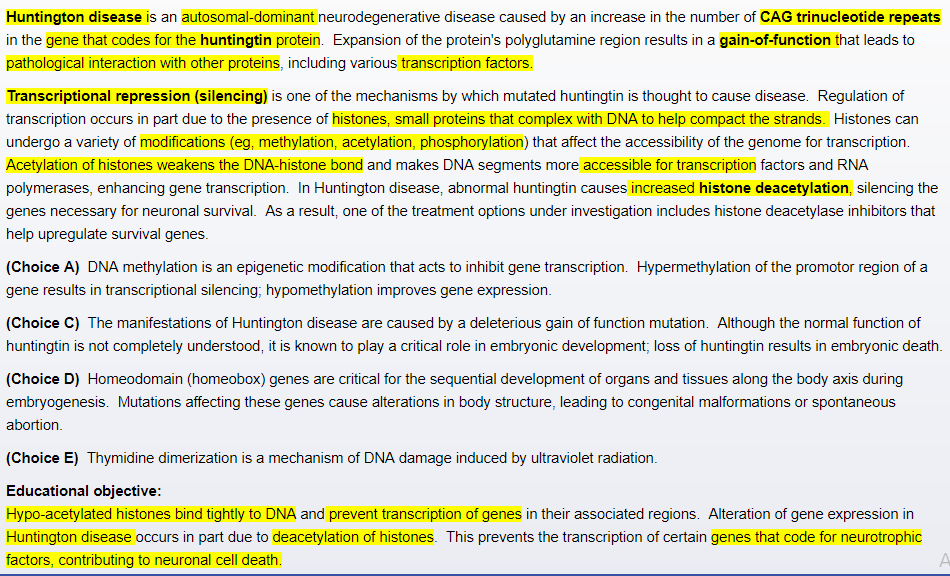
*Acetylation of DNA
- Activates DNA
- deactelyation = deactivates DNA
--> seen in HD
Pathophys of HD
- CAG repeats --> cause deacetylation --> trasncription silencing
*Mitochondria
Mitochondria
example
Example:
Notes:
- note when looking for mitochondria on electron microscope, first identify is there is a nucleus and a nucleosome present, then find the mitochondria which have lines that make up their matrix
*Peroxisomes
- PER - OXIDATION = fatty acid oxidation that can't be done by the mitochondria
--> VLCFA = very long chain fatty acid oxidation in the peroxisome
*Nucleosome
- contained in the nucleus
- main site for rRNA = ribosomal RNA synthesis and assembly before they are sent out
- nucleus has 2 main jobs
--> keep DNA + make ribosomes to make the proteins
Mitochondria
example
Example:
Notes:
- note
*Peroxisome Disorders
- can't break down VLCFA = very long chain fatty acid oxidation in the peroxisome
NUCLEAR pot transcription modification of pre mRNA
- 3 major steps of postranscription modification
*5' Cap
- 1/3 nuclear step of Post-transcriptional mRNA modification
- stops the mRNA degraded in the nucleus
*3' Poly A Tail
- 2/3 nuclear step of pre mRNA modification
- stops the mRNA degraded in the cytoplasm
--> keep losing the 3' tail
--> this makes sense as you need a STOP at the end of RNA, this is more important
*Splicing out of introns
- LASTS = 3/3 nuclear step of pre mRNA modification
*Introns and exons
- EXONS = Actual mRNA that get EXPRESS
- INTRON = just there to INTERRUPT the ACTUAL mRNA
*PCR
(Polymerase Chain Reaction)
*ELISA kits
ELISA kits
- Enzyme-linked immunosorbent Assay (direct / indirect)
- for identifying antibodies and proteins
Example:
Notes:
- to identify whether someone has been exposed to a certain virus and has antibodies / immunoglobulins to that virus
- step 1 - line wells with viral antigen and add patient serum to the wells
--> if they have antibodies to the virus they will bind to the antigens of the well - step 2 - wash the plate
--> this removes any other proteins etc. that have not binded to the specific antigens - step 3 - add the substrate-modifying E = EL part of ELISA
--> the enzyme that is linked to a specific substrate that will activate the enzyme is key to the whole process of the ELISA kit
--> the enzyme here is attached to an anti-human immunoglobulin antibody
--> this attaches to the patient's human antibody - step 4 wash again
- step 5 - add the substrate to the wells to activate the enzyme
- the degree of colour change or whatever the enzyme does will tell you how much of the antibody is present in the well
--> the substrate linked enzyme = substrate-EL is usually peroxidase
Notes:
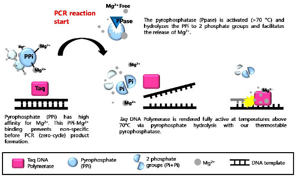
- "PCR = 95.7 is a TAQy station that PRIMES DNA for NEW TRIAL songs"
- PCR 95.7
--> 95 degrees C = denature the original
--> 55 degrees C = cools down for primer to attach to the needed sequence
--> 72 degrees C = TAQ polymerase elongates the new DNA strand - TAQy station
--> TAQ polymerase = thermos aquatic polymerase is able to work at high temps of 72 degrees - PRIME DNA for NEW TRIALS
--> 3 ingredients needed for PCR are: primer, DNA original, nucleotide triphosphates
*Agglutination inhibition tests
- AI tests have antigens to something like beta hCG
- add urine/solution to the AI fluid
- then you add latex coated in hCG (or what yu are testing for)
- agglutination happens with latex coated = negative result
--> since there was no hCG before - NO agglutination with latex coated = positive result
--> since there is hCG present from before
Triple alpha helix in CAG COLL - A - GEN
- CAG COLL - A - GEN
--> C = Vitamin C neded for collagen synthesis
--> A = alpha triple helices
--> G = glycine every 3 AAs gives the flexible structure for collagen / with prolines in between that give the rigidity needed - note the triple helix structure is ONLY in the PROCOLLAGEN polypeptide that has to be cleaved
- PROCOLLAGEN is then cleaved into collagen fibrils that all CROSS LINKED to each other by LYSYL OXIDASE
--> think of collagen as being strong like a bacterial cell wall because it has individual building blocks = collagen fibrils that are crosslinked to gether to make it strong
--> just like in bacterial cell wall the D-ALA - D ALA are crosslinked to make a strong bacterial cell wall
*Transcription factors
- leucine zipper transcription factor
*leucine zipper - Transcription factors
- leucine zipper transcription factor
*Transport across membranes
- passive diffusion vs active protein carrier transport
Clinical Cases
Clinical Case
Clinical Case
Notes:
- note that
Reading mRNA Practical Example
- mRNA is read in the 5' --> 3' direction
- opposite to DNA reading
- think the AAs need to match the original DNA so the mRNA is read in the opposite direction to the DNA
Clinical Case
Notes:
- note that tRNA has a CCA end at the 3' end
--> CCA has OH group attached to it where the AAs bind - tRNA is held together by hydrogen bonds of complement sequences
- has a DAVENPORT clover LOOP structure
- D = D loop
- A = anticodon loop
- V = small V loop
T = T loop
Charged vs. Uncharged tRNA
- Uncharged tRNA goes out to bind to AAs
- once tRNA binds to AAs it becomes charged and can bind to the A site of the Ribosome for giving its AA
8releasing factors
- bind to the ribosome and casue the release of the polypeptide when the mRNA reaches one of the 3 STOP codons
--> UAA
--> UGA
--> UAG
*Flanking region
- note you need to know the nucleotide sequence of the 2 flanking regions on either side of the exon or gene you want to copy and amplify
- the flanking region = DNA primer binding region
Clinical Cases
Clinical Case
Clinical Case
Notes:
- note that
Mechanics of splicing
- "Get U!" -- INTRONS -- "All GONE!"
--> GU -- INTRONS -- AG - GU marks the 5' end of introns to START the cut by snRNPs and the splicosome
--> snRNPs = small nuclear ribonucleic Proteins - AG marks the 3' end of introns to END the cut by snRNPs and the splicosome
*Methylation of DNA
- Methylation of cytosine or adenosine in DNA
--> "makes DNA MUTE"
--> expecially methylation of CpG islands in DNA - Methylation of Histones though
--> "MAY make DNA MUTE or it MAY not"
--> this is the exception to the rule
*FISH = Flourescant In Situ Hybridization
- this is a lab technique for highlighting chromosome abnormalities
--> this is the even smaller version of the SNW DRP lab tests
Telomeres and telomerase in Malignancy
Clinical Cases
Clinical Case
Clinical Case
Notes:
- note that
Telomeres and telomerase in Malignancy
Notes:
- note that only germline cells have telomerase naturally since they are dividing so often and need telomerase to make and replace the telomere
- other than this telomerase activity is pathogenic and is present in 90% of cancers since telomeres normally signal through the p53 tumor supressor gene to start apoptosis when DNA has undergone too many divisions
Case example:
*Telomerase enzyme
- telomerase adds TTAGGG repeats at the 3' ends of DNA
--> these end pieces = TAGs are telomeres - prevent them from being degraded
- this is mostly done in the stem cells of the body since they need much longer telomeres
- other mature cells of the body have shorter telomeres
--> when a telomere reaches a certain short length it signals for apoptosis of that cell
Clinical Cases
Clinical Case
Clinical Case
Notes:
- note that
*Lyonization = X inactivation
- in each cell of the body of a female, one of the X chromosomes is randomly chosen for inactivation
--> becomes a Barr Body
--> done by heavy Methylation = MUTED DNA
Nucleotide Excision Repair
- similar to BASE excision and GEL PLease
- glycosylase , endonuclease, lyase, polymerase, lygase
- key is there is no Glycosylase since you are not taking out and switching the base
- this is from UV damage and repairs dimerized pyrimidines
--> thymine and cytosine
CYTOPLASM *PBODY modification
- use microRNA to degrade or change the mRNA
*Endoplasmic Reticulums and Golgi Apparatus
-
*Rough ER
- has ribosomes and makes proteins
*Smooth ER
- has NO ribsomes and is used for making steroid hormones
- thus high in androgen making cells, gonads, etc.
*Golgi Apparatus
- has no ribosomes and is used to packege proteins into secretory vessicles for transport from the cell


